|
Abstract Cerebral microbleeds (CMBs) are small haemorrhages nearby blood vessels. They have been recognized as important diagnostic biomarkers for many cerebrovascular diseases and cognitive dysfunctions. In current clinical routine, CMBs are manually labelled by radiologists but this procedure is laborious, time-consuming, and error prone. In this paper, we propose a novel automatic method to detect CMBs from magnetic resonance (MR) images by exploiting the 3D convolutional neural network (CNN). Compared with previous methods that employed either low-level hand-crafted descriptors or 2D CNNs, our method can take full advantage of spatial contextual information in MR volumes to extract more representative high-level features for CMBs, and hence achieve a much better detection accuracy. To further improve the detection performance while reducing the computational cost, we propose a cascaded framework under 3D CNNs for the task of CMB detection. We first exploit a 3D fully convolutional network (FCN) strategy to retrieve the candidates with high probabilities of being CMBs, and then apply a well-trained 3D CNN discrimination model to distinguish CMBs from hard mimics. Compared with traditional sliding window strategy, the proposed 3D FCN strategy can remove massive redundant computations and dramatically speed up the detection process. We constructed a large dataset with 320 volumetric MR scans and performed extensive experiments to validate the proposed method, which achieved a high sensitivity of 93.16% with an average number of 2.74 false positives per subject, outperforming previous methods using low-level descriptors or 2D CNNs by a significant margin. The proposed method, in principle, can be adapted to other biomarker detection tasks from volumetric medical data. |
|
Results |
||
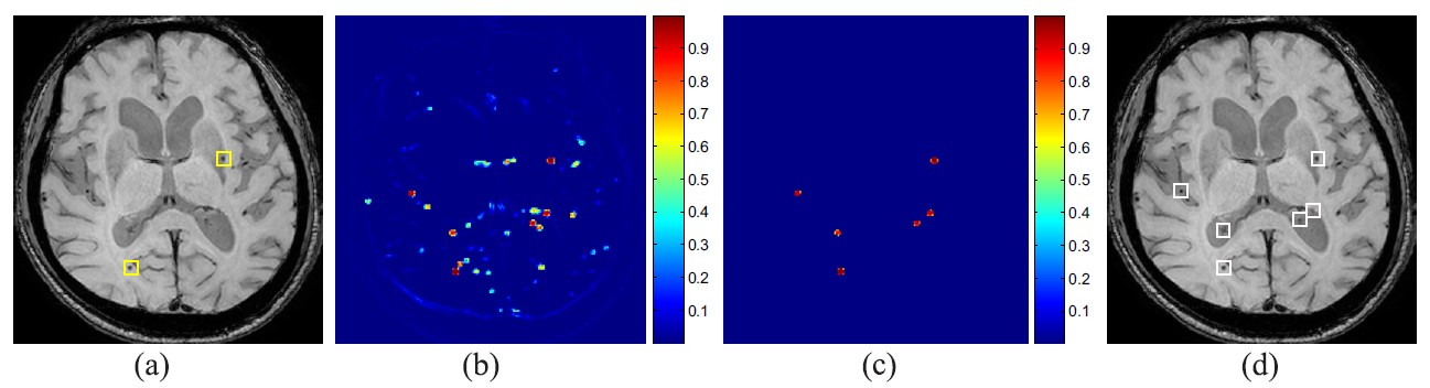
Fig. 1. Typical results of the 3D FCN screening model witSh score volume projection onto the axial plane. (a) Raw data with true CMBs (yellow rectangles). (b) 2D projection of the score volume generated with FCN. (c) 2D projection of the post-processed score volume. (d) Retrieved candidates (white rectangles). |
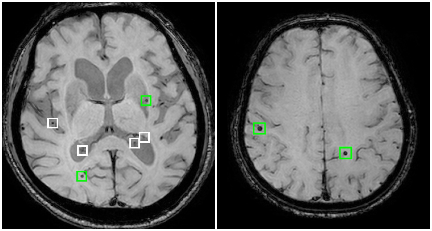
Fig. 2. Examples of CMB detection results (viewed in axial planes). Green rectangles denote the correctly detected CMBs and white rectangles denote the removed false positive candidates by our method. |
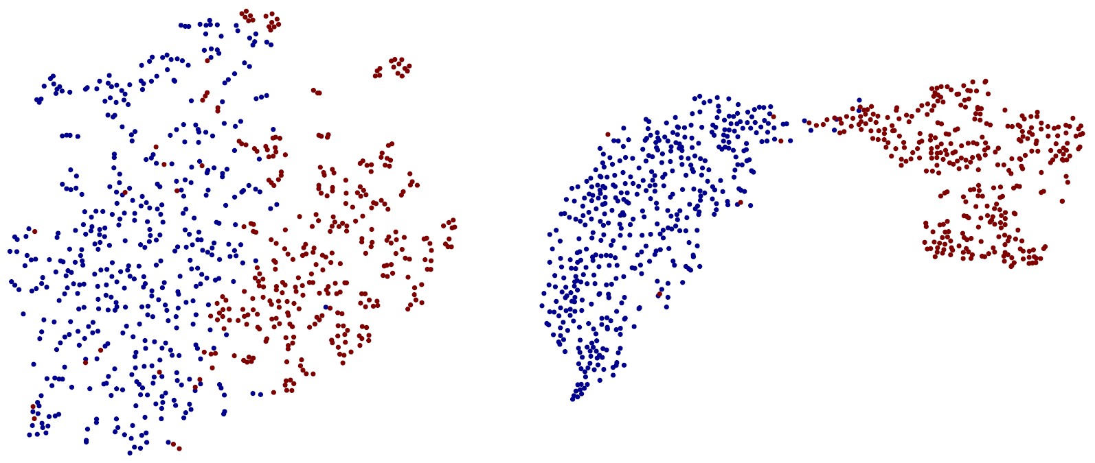
Fig. 3. Feature embedding from 2D-CNN-SVM (left) and 3D CNN methods (right) with t-SNE toolbox. The red and blue colors correspond to the CMBs and non-CMBs, respectively. |
Downloads |
 |
Q. Dou*, H. Chen*, L. Yu, J. Qin, L. Shi, P. A. Heng, et al. "Automatic Detection of Cerebral Microbleeds from MR Images via 3D Convolutional Neural Networks." |